In short
Diabetic foot osteomyelitis is an infection of the bone that usually spreads from a foot ulcer sitting over bone. Clues include an ulcer that will not heal, bone felt with a sterile probe, a red swollen toe and raised inflammation markers in a blood test. It is diagnosed with an X-ray, often an MRI scan, and ideally a bone sample. Treatment is antibiotics, sometimes with surgery to remove infected bone.
Diabetic foot osteomyelitis is an infection of a bone in the foot, including the bone marrow, in someone with diabetes.1 It usually happens when bacteria spread straight from an ulcer into the bone underneath, rather than arriving through the bloodstream.2 Bone infection is one of the more serious kinds of diabetic foot infection, because it greatly raises the risk of minor and major amputation.2 This article explains how it is suspected, confirmed and treated.

What causes diabetic foot osteomyelitis?
Nerve damage (neuropathy) means you may not notice a wound, and poor circulation slows healing. When an ulcer gets deep, germs can reach the hard outer layer of the bone (the cortex) and then the marrow inside. The IWGDF/IDSA guideline uses the single word osteomyelitis for both, because they are hard to tell apart and are treated the same way.2
The NHS lists diabetes as a key risk factor, especially if you have a foot ulcer.3 In the IWGDF/IDSA grading system, bone infection is shown by adding '(O)' to a moderate or severe grade, for example 3(O).2
Symptoms and clues of bone infection in the foot
Bone infection can be quiet. Because of nerve damage there may be little pain, and the skin may look only a little inflamed. The guideline lists signs that should make your care team think of it2:
- an ulcer that has been there for many weeks, or one that is wide or deep
- an ulcer over a bony part of the foot, or one where bone can be seen
- a red, swollen 'sausage' toe
- bone felt at the bottom of the wound with a sterile probe
- raised blood markers of inflammation, such as ESR or CRP
NICE, which sets guidance for the NHS, advises considering osteomyelitis in anyone with diabetes who has a local infection, a deep foot wound or a long-standing (chronic) foot wound. It also warns that bone infection can be there even when blood tests, X-rays or the probe-to-bone test are normal.4
What is the probe-to-bone test?
Your doctor or podiatrist gently passes a sterile, blunt metal probe into the wound. If it touches a hard, gritty surface, the test is positive.2 It is quick, inexpensive and practically harmless.
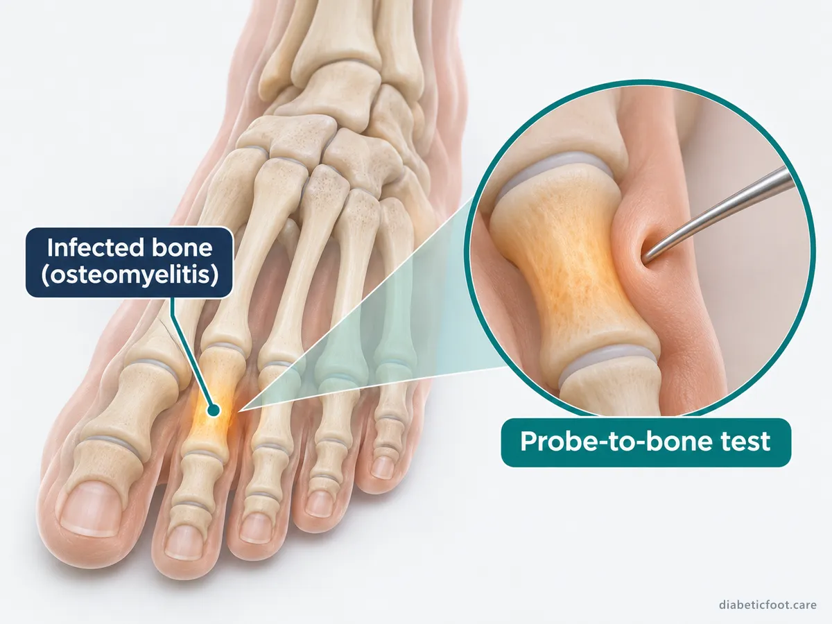
The test is most useful alongside other findings. A positive result in a high-risk wound points towards bone infection. A negative result in a low-risk wound helps rule it out.2 How reliable it is depends on the experience of the person doing it, and different examiners only moderately agree.2
X-ray and MRI for diabetic foot osteomyelitis
| Test | Role |
|---|---|
| Plain X-ray | First imaging test; may show loss of bone outline, erosion, a reaction of the bone lining (periosteum) or fragments of dead bone |
| Repeat X-ray | Considered after 2 to 3 weeks if the first X-rays are normal but suspicion stays high |
| MRI scan | Used when the diagnosis is still in doubt after examination, X-ray and blood tests |
| PET, white cell scan or SPECT | Alternatives when MRI is not possible |
| Bone sample | Confirms infection and identifies the germ |
These tests follow the IWGDF/IDSA guideline.2 X-rays are cheap and easy to get but miss early infection, which is why a normal X-ray may be repeated.2 NICE also advises considering an MRI scan if osteomyelitis is suspected but the first X-ray does not confirm it.4
Why a bone biopsy or bone sample helps
A bone sample is the accepted standard test for diagnosis. It is the only sure way to identify the germs in the bone.2 It can be taken during an operation or with a needle through the skin (percutaneously). The guideline suggests taking bone samples, rather than soft tissue samples, when osteomyelitis in the diabetic foot is suspected.2
This matters because cultures from the wound surface and from the bone often differ. Most studies report that they agree less than half the time.2 Knowing the real germ helps your team choose an antibiotic that works without being broader than needed. A bone biopsy is not always available or suitable, and your team will weigh up the benefits against the small risks.
Osteomyelitis or Charcot foot?
Both can cause a red, warm, swollen foot. Charcot foot (Charcot neuro-osteoarthropathy) can also cause X-ray changes that look like bone infection.2 NICE advises suspecting acute Charcot foot when there is redness, warmth, swelling or a change in the foot's shape, particularly when the skin is unbroken, and especially if you have nerve damage.4
An open ulcer that reaches the bone points more towards infection. Unbroken skin points more towards Charcot foot. In difficult cases an MRI scan, and sometimes a bone sample, are needed. Read more about Charcot foot and diabetes.
How is bone infection in the foot treated?
The IWGDF/IDSA guideline describes two main approaches to treating diabetic foot osteomyelitis, and the choice is made for each person2:
- Antibiotics without surgery can be considered for bone infection in the front of the foot (forefoot). This applies when there is no urgent need to drain infection, no peripheral artery disease (narrowed leg arteries) and no exposed bone. For carefully chosen people, studies suggest results similar to surgery.2
- Surgery to remove infected bone, plus antibiotics is considered in other situations, especially with widespread soft tissue infection, exposed bone or dead bone.2
In the guideline, antibiotics typically last 6 weeks when no bone is removed. They last up to 3 weeks when infected bone is left at the edge after a minor amputation, and only a few days when all the infected bone has been removed.2 Many people can take tablets, so bone infection alone does not always mean a hospital stay.2 If blood flow is very poor, restoring it is part of treatment.2 For how antibiotics are chosen and reviewed, see our guide to diabetic foot infection treatment.
Remission and outlook
Diabetic foot osteomyelitis settles slowly and can come back, so doctors talk about remission. This means there is no ongoing or new infection in the same place. The guideline advises judging remission at least 6 months after antibiotics stop, and longer follow-up has traditionally been used.2 Falling blood markers, and X-rays showing no further bone damage, are reassuring signs.2
If the pressure that caused the original ulcer is not dealt with, infection may come back in the same place.2 When bone or soft tissue cannot be saved, part of the foot may need to be removed. Our guide to diabetic foot gangrene and amputation risk explains this.
Frequently asked questions
What are the symptoms of osteomyelitis in a diabetic foot?
There may be few symptoms. Clues include an ulcer that has not healed after several weeks, an ulcer over a bony point or with bone showing, a red, swollen 'sausage' toe, discharge and raised inflammation markers in blood tests. Because nerve damage can hide pain, a painless wound can still involve the bone. Any deep or long-standing wound should be checked by your foot team.
Can diabetic foot osteomyelitis be treated without surgery?
Sometimes. Antibiotics alone can be considered for bone infection in the front of the foot when there is no urgent need to drain pus, no significant artery disease and no exposed bone. In other cases, removing the infected bone is usually advised, followed by a shorter course of antibiotics. Your team will explain which approach suits your foot and your general health.
Does an X-ray always show bone infection?
No. An X-ray is the first test, but it often looks normal early on, because bone changes take time to show. If your team is still concerned, the X-ray may be repeated after 2 to 3 weeks, or an MRI scan arranged. Charcot foot can also cause X-ray changes, so results are read together with the examination and blood tests.
Why might I need a bone biopsy?
A bone sample is the most reliable way to confirm diabetic foot osteomyelitis and find out which germ is causing it. Samples from the wound surface often do not match the germs in the bone, which can lead to the wrong antibiotic. A biopsy may be taken during surgery or with a needle through the skin. It helps your team choose a targeted antibiotic and avoid treatment that is broader than needed.
Can bone infection in the foot come back?
Yes, diabetic foot osteomyelitis can return. That is why doctors talk about remission and keep seeing people for at least 6 months after antibiotics stop. Taking pressure off the foot, wearing suitable footwear, keeping your blood glucose steady and going to regular foot reviews all help lower the risk of a new ulcer and a new infection in the same place.
If you have signs of infection now — redness spreading, a hot swollen foot, discharge or feeling unwell — contact your GP practice, foot protection service or NHS 111 today, or call 999 if you are seriously unwell. Do not wait to contact us.

Specialist assessment in Istanbul
Would you like a specialist opinion?
Assoc. Prof. Dr. Hüsrev Diktaş is an infectious diseases and clinical microbiology specialist in Istanbul with a clinical focus on diabetic foot infections. International patients can ask about an assessment directly.
References
- International Working Group on the Diabetic Foot. Definitions and criteria for diabetes-related foot disease (IWGDF 2023 update). 2023. https://iwgdfguidelines.org/wp-content/uploads/2023/05/DMRR3654.pdf
- International Working Group on the Diabetic Foot / Infectious Diseases Society of America. Guidelines on the diagnosis and treatment of foot infection in persons with diabetes (IWGDF/IDSA 2023). 2023. https://iwgdfguidelines.org/wp-content/uploads/2023/07/IWGDF-2023-04-Infection-Guideline.pdf
- NHS. Osteomyelitis. 2023. https://www.nhs.uk/conditions/osteomyelitis/
- National Institute for Health and Care Excellence. Diabetic foot problems: prevention and management (NG19), recommendations. 2015, updated 2019. https://www.nice.org.uk/guidance/ng19/chapter/Recommendations
This page gives general information for people in the UK. It is not a diagnosis and does not replace advice from your own GP, diabetes team or foot protection service. Medical disclaimer · How we write our content